A MULTIFRACTAL BRAIN
Perhaps the most exciting aspect of this hypothesis is that multifractal tensegrity extends into the brain. The brain is most often imagined as being isolated from the rest of the body, afloat in a vault of magical membranes and fluids. Turvey & Fonseca highlight the possible role of a lesser known mechanical connection between the muscles of the upper neck and the membranes which envelop the spinal cord and brain.

This connection from the multi fractal tensegrity of the body into the membranous structure surrounding the brain opens up the possibility of a tensional interaction between the ECM-fibroblast network of the body and the brains’ own specialised hyaluron-astrocyte network.
Fibroblasts secrete ECM into every space of the body – there are no empty spaces. Similarly the brain is invested with supportive glial cells, including astrocytes – there are no empty spaces in the brain either.
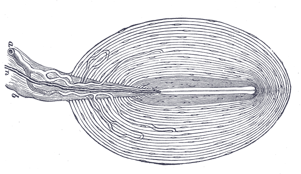
This maximally packed and interconnected architecture, of the body and brain, is exemplified by the onion-like Pacinian corpuscle (a mechanoreceptive nerve ending in the connective tissue of the body). The concentric lamellae, surrounding the unmyelinated nerve fibres at the corpuscle’s core, are thinly separated – ECM fills these spaces.

This neural architecture is mirrored in the brain – the narrow spaces between neurons are filled with ECM. This specialised ECM (not the same as the body’s ECM) is secreted by neurons and glial cells. Until quite recently, the prevailing understanding was that the close packing of neurons and glial cells left no room for any other material. Newer technology has shown that empty spaces are filled with a soup of ECM molecules. It is now known that ECM accounts for 10–20% of mass in a mature brain. The ECM of the brain is very different from that of the body. It is synthesised by and embedded with various types of glial cells – brain ECM is predominantly composed of the glycosaminoglycan called hyaluronan.
THE BRAINS ECM
Relative to its mass – hyaluronan occupies a large volume. Even at low concentrations hyaluronan forms gels. Hyaluronan gels bind large quantities of water, this water is extruded when compressed and replenished when compression is released. The inward drawing of water creates a swelling pressure which enables the brain’s ECM to withstand compressive forces.
Hyaluronan gels provide the brain with a capacity to tolerate compression – one side of the biotensegrity equation. The opposing side of the biotensegrity equation is tension, this is also fulfilled by hyaluronan which has been shown to exhibit multiple structural conformations (including extended chains or cables, helical coils, and toroids). These structural conformations are resistant to tension.
ASTROCYTES
Hyaluronan gels embed both neuronal and non-neuronal cells. Non-neuronal cells, including astrocytes and hyaluronan gels define the brain’s connective tissue network. The brain’s connective tissue network is enveloped by, and continuous with, the brains collagen-based meningeal membranes. The brains collagen-based meningeal membranes are part of the MCS. This forms an interface between the body and the brain.
Astrocytes have a critical role in merging the tensional networks of the body and brain. Astrocytes are highly mechano-sensitive glial cells, with numerous long processes.

This enables multiple connections with neurites (the axons and dendrites of neurons), blood vessels, other astrocytes and the inner layer of the meninges, the pia mater. The three-dimensional interconnectedness of astrocytes is ideal for sensing mechanical disturbances. Mechanical disturbances include the ongoing adjustment of neurites as they maintain steady tensional and spatial relationships. Mechanical disturbances of the meninges, which accompanies all movement and actions of the body, are sensed by the astrocyte network. These similarities and continuities with the rest of the body make it clear why the brain and spinal cord have been included in the multi fractal tensegrity.

INTERCONNECTIONS OF NEURAL, NON-NEURAL AND VASCULAR ELEMENTS IN THE BRAINS ECM
Turvey and Fonseca state:
‘If the possibility proved to be actuality, then a reasonable expectation would be that the interplay of tension and compression from near and distal parts of the MCS system are communicated to cranial dura mater and thence to cranial pia mater and the brain’s connective tissue—the hyaluronon-based ECM and the various cells, neurons and glia, embedded within it. By the same token, there should be a reciprocal consequence, however minimal, for tension and compression arising within the brain’s connective tissue. Astrocyte stress-fibers in vitro organize into parallel bundles that can span many cells suggesting that the astrocyte network can exert long-range forces on the ECM. If the impedance matching was of the right order, then such forces could, in principle, modulate the tensile state of the pia mater, and communicate subtle mechanical states of brain tissue to the multifractal tensegrity in the large’.
This arrangement has the possibility of influencing tensional relationships of the pia mater (the most intimate dural layer surround the brain and CNS). This tensional relationship has the potential to influence the ’brain-wide pathway of waste clearance’ referred to as the glymphatic system. It has been shown that non-neuronal cells like astrocytes play a vital role in neurobiological health and intrinsic brain function.
THE HAPTIC MEDIUM – AN ORGAN OF PERCEPTION
The haptic medium, in the language of ecological psychologist J.J Gibson, can be described as an organ of perception. As far back as 2003, Robert Schleip was suggesting that fascia might turn out to be ‘our most important perceptual organ’ particularly as it is the source of proprioceptive, visceral and interoceptive sensation.
As discussed previously, fascia is richly innervated, possessing ten times the quantity of sensory nerve receptors as muscle. More recently fascia has been shown to possess a rich network of autonomic motor nerves. By adding together the number of mylinated proprioceptors and free nerve ending in fascia, we find that this total is greater than the retina which is considered our richest sensory organ.
With further research the fascial body is likely to emerge as our most significant sensory organ. This fascial body encompasses not only the fascial network or soft tissue ectoskeleton but also the many million endomysial sacs and membranous pockets whose total surface area far surpasses that of the skin or any other organ – the fascial body seemingly also encompasses the brain.
FEATURED IMAGE:
H.V. Carter. Anatomy of the human body (1918).
